Current: Investigating the Neuro-Immune Interaction in the Brain:
This project is centered around the intriguing exploration of how the brain’s immune system can influence behavior and remodel neuronal structures. To study this, we employ cutting-edge microscopy technology for automated quantification of morphological structures. Our goal is to unravel the intricate ways in which neural circuits may be altered when the immune system is activated or suppressed. Through this research, we aim to shed light on the complex interactions between the brain’s immune response and its impact on brain function, opening doors to potential therapeutic applications and a deeper understanding of neurological conditions.
Above: Postnatal Developmental Maturation Trajectory of Microglia (immune cells in the brain).
Current: Role of Environmental Enrichment in Learning and Memory
This project focuses on how the quality of environment can impact our ability to learn and remember skills. By providing enriching environments, we aim to understand how these positive experiences can boost our learning and memory capacities. Through careful research and analysis, we hope to uncover the underlying mechanisms behind this phenomenon, which could have practical implications for improving cognitive functions in general. This will be mainly emphasizing on the anatomical indicators of structural plasticity, namely, dendritic spines, while correlating with behavior, and manipulation of specific circuits in the brain.

Above: Using dendritic spines as an anatomical proxy of structural plasticity at the synaptic level. A: Golgi-impregnated dendritic segment in a cortical pyramidal neuron (S1 Layer 4 Spiny Stellate Cell, 60X). B: 2D Rendering of the same dendritic segment in A, color codes correspond to different spine morphology indicated in C. C: Quantification of spine categories based on physical length from tip to base of dendritic spine. D: Immunofluorescent staining revealing spine-specific proteins (e.g., spinophillin) colocalized with spine heads (GPF-labeled).
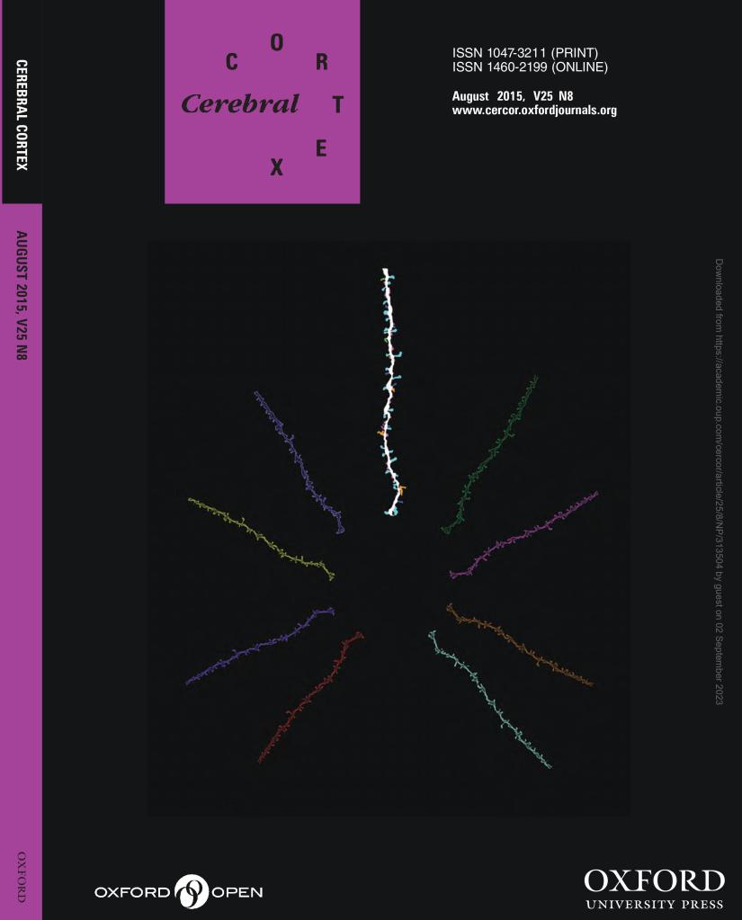
Above: Dendritic segment detailing different morphology of spines. Appeared as cover image at Cerebral Cortex (August 2015). See Chen et al., 2015.
